


Epidural & Spinal Anesthesia: Understanding the Facts
Women's Health & Education Center's Contribution. We hope that you, the reader, will benefit from this symposium on pain management during labor and delivery, and we trust that it will improve your understanding.
Epidural & spinal anesthesia (regional anesthesia) has become more popular recently, because they are well suited to pain management during labor. It offers the most effective form of pain relief and is used by most women in the United States. There are no absolute indications for epidural anesthesia. However, there are clinical situations in which patient preference, patient physiology, or the surgical procedure makes epidural anesthesia the technique of choice. Thus, epidural anesthesia is an indispensable part of modern anesthetic practice, and every anesthesiologist should be adept at performing this procedure. Proficiency in epidural anesthesia requires a thorough understanding of the anatomy of the spine and spinal cord. Epidural anesthesia can be used to extend analgesia into the postoperative period or to provide analgesia to non-surgical patients. Spinal and epidural anesthesia each have advantages and disadvantages that may make one or the other technique better suited to a particular patient or procedure.
The purpose of this document is to understand indications, contraindications, techniques and areas of future research in regional anesthesia (epidural and spinal anesthesia). In obstetric patients, regional analgesia refers to a partial to complete loss of pain sensation below the T8 to T10 level. In addition, a varying degree of motor blockade may be present, depending on the agents used. The spine consists of 33 vertebrae (7 cervical, 12 thoracic, 5 lumbar, 5 fused sacral, and 4 fused coccygeal). The vertebral bodies are stabilized by five ligaments that increase in size between the cervical and lumbar vertebrae. In most obstetric patients, the primary indication for epidural analgesia is the patient's desire for pain relief. The American Society of Anesthesiologists (ASA) and the American College of Obstetricians and Gynecologists (ACOG) recommend that third-party payers should not deny reimbursement for regional analgesia and anesthesia because of an absence of other medical indications.
Anatomy of Epidural Space:
The epidural space is the space that lies between the spinal meninges and the sides of the vertebral canal. It is bounded cranially by the foramen magnum, caudally by the sacrococcygeal ligament covering the sacral hiatus, anteriorly by the posterior longitudinal ligament, laterally by the vertebral pedicles, and posteriorly by both the ligamentum flavum and vertebral lamina. The epidural space is not a closed space but communicates with the paravertebral space by way of the intervertebral foramina. The epidural space is shallowest anteriorly where the dura may in some places fuse with the posterior longitudinal ligament. The space is deepest posteriorly, although the depth varies because the space is intermittently obliterated by contact between the dura matter and ligamentum flavum or vertebral lamina. Contact between the dura mater and the pedicles also interrupt the epidural space laterally. Thus, the epidural space is composed of a series of discontinuous compartments that become continuous when the potential space separating the compartments is opened up by injection of air or liquid (1).
The spinal meninges consist of three protective membranes (dura mater, arachnoid mater, and pia mater) that are continuous with the cranial meninges. The dura mater is the outermost and thickest meningeal tissue. The inner surface of the dura mater abuts the arachnoid mater. There is potential space between these two membranes called the sub-dural space. Occasionally, drug intended for either the epidural space or the subarachnoid space is injected into the sub-dural space. Sub-dural injection has been estimated to occur in 0.82% to 10% of intended epidural injections. The arachnoid mater is a delicate, avascular membrane composed of overlapping layers of flattened cells with connective tissue fibers running between the cellular layers. The spinal pia mater is adherent to the spinal cord and is composed of a thin layer of connective tissue cells interspersed with collagen. The pia mater also gives rise to the dentate ligaments, which are thin connective tissue bands extending from the side of the spinal cord through the arachnoid mater to the dura mater. These ligaments serve to suspend the spinal cord within the meninges.
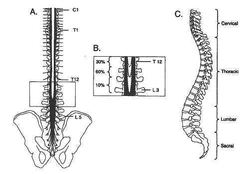
(A) Posterior and (C) Lateral views of the human spinal column. The inset (B) depicts the variability in vertebral level at which the spinal cord terminates.
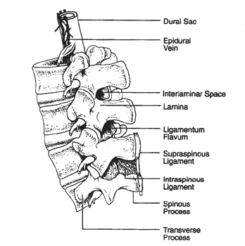
Details of the lumbar spinal column and epidural space. The epidural veins are largely restricted to the anterior and lateral epidural space.
Techniques:
Identifying individual vertebrae is important for correctly locating the desired interspace for epidural and spinal blockade. The spine of C7 is the first prominent spinous process encountered while running the hand down the back of the neck. The spine of T1 is the most prominent spinous process and immediately follows C7. The 12th thoracic vertebra can be identified by palpating the 12th rib and tracing it back to its attachment to T12. A line drawn between the iliac crests crosses the body of L5 or the L4-5 interspace. Midline insertion of an epidural needle is least likely to result in unintended meningeal puncture. Spinal and epidural anesthesia should be performed only after appropriate monitors are applied and in a setting where equipment for airway management and resuscitation are immediately available. Before positioning the patient, all equipment for spinal block should be ready for use (e.g. local anesthetics mixed and drawn, needles uncapped, prep solution available). Spinal and epidural needles are named for the design of their tips. Epidural needles have a larger diameter than spinal needles to facilitate the injection of fluid or air when using the "loss-of-resistance". A catheter is placed in the epidural space, allowing for continuous epidural infusion of local anesthetic agents or narcotics. The advantage of this method is that medication can be titrated over the course of labor as needed. In addition, epidural catheters placed for labor analgesia can be used for cesarean delivery or postpartum tubal ligation. Modern epidural preparations that combine a low-dose local anesthetic, such as bupivacaine, levobupivacaine, or ropivacaine, with an opioid agonist are preferred because they decrease motor blockade and result in an increased rate of spontaneous vaginal delivery (2).
Careful attention to patient positioning is critical to successful spinal puncture. Using the iliac crests as a landmark, the L2-L3, L3-L4 and L4-L5 interspaces are identified and the desired interspace is chosen for needle insertion. Interspaces above L2-L3 are avoided to decrease the risk of hitting the spinal cord with the needle. Penetration of the dura mater produces a subtle "pop" that is most easily detected with the pencil-point needles. Single-shot spinal anesthesia provides excellent pain relief for procedures of limited duration, such as cesarean delivery, the second stage of labor, rapidly progressing labor, and postpartum tubal ligation. A long-acting local anesthetic often is used, with or without an opioid agonist. The duration of anesthesia is approximately 30-250 minutes depending on the drugs used. However, because of its inability to extend the duration of action, single-shot spinal analgesia is of limited use for the management of labor.
Combined Spinal Epidural:
Combined spinal-epidural anesthesia (CSEA) is a useful technique by which a spinal block and an epidural catheter are placed simultaneously. This technique is growing in popularity because it combines the rapid onset, dense block of spinal anesthesia with the flexibility afforded by an epidural catheter. There are special epidural needles with a separate lumen to accommodate a spinal needle available for CSEA. The spinal component of CSEA may be an intrathecal narcotic plus a small amount of a local anesthetic. Failure of the spinal component occurs at a rate of 4% with CSEA, but the block can be supplemented with epidural catheter (3). Although CSEA shows great promise, additional, prospective studies are necessary to identify the relative risks and limitations of the technique.
Absolute Contraindication to Regional Anesthesia:
The absolute contraindication to spinal or epidural anesthesia is patient refusal. However, several pre-existing conditions increase the relative risk of these techniques, and the anesthesiologist must carefully weigh the expected benefits before proceeding. Some conditions that increase the risks are:
- Hypovolemia or shock increases the risk of hypotension.
- Maternal coagulopathy or thrombocytopenia increases the risk of epidural hematoma. Patients with platelet counts of 50,000-100,000/ µL may be considered potential candidates for regional anesthesia.
- Skin infection over the site of needle placement. Sepsis increases the risk of meningitis.
- Pre-existing neurologic disease, particularly diseases that wax and wane (e.g. multiple sclerosis).
- Increased intracranial pressure increases the risk of brain herniation when CSF is lost through the needle, or if a further increase in intracranial pressure follows injection of large volumes of solution into the epidural or subarachnoid spaces.
Complications:
- Backache although postoperative backache occurs after general anesthesia, it is more common after epidural and spinal anesthesia. Compared with spinal anesthesia, back pain after epidural anesthesia is more common (11% vs. 30%) and of longer duration. The etiology of backache is not clear, although needle trauma, local anesthetic irritation, and ligamentous strain secondary to muscle relaxation have been offered as explanations.
- Post-dural Puncture Headache (PDPH) it is a common complication of spinal anesthesia, with reported incidence as high as 25% in some studies. The risk is less in epidural anesthesia, but it occurs in up to 50% of young patients after accidental meningeal puncture. PDPH usually resolves spontaneously in few days to a week for most patients. Initial treatment is appropriately conservative if this meets the patient's needs. Bed rest and analgesics is necessary and the mainstay of conservative treatment. Caffeine has also been shown to produce short-term symptomatic relief. Epidural blood patch is an alternative for patients who are unable or unwilling to await spontaneous resolution of PDPH (4).
- Hearing loss a transient (1-3 days) mild decrease in hearing acuity (>10 db) is common after spinal anesthesia patients with an incidence of roughly 40% and a 3:1 female: male predominance (5). The mechanism of hearing loss in these studies is unclear, but the marked female predominance, the absence of PDPH, and the difference in incidence between prilocaine and bupivacaine suggests that CSF leak is not the cause.
- Neurologic Injury serious neurologic injury is a rare but widely feared complication of epidural and spinal anesthesia. Multiple large series report the incidence in ~0.03-0.1% of all central neuraxial blocks, although in most of these series the block was not clearly proven to be causative. Local anesthetic intended for epidural and intrathecal use can themselves be neurotoxic in concentrations used clinically. The mechanism by which local anesthetics produce cauda equine syndrome in not yet clear; however, the evidence suggests that local anesthetics produce toxic damage by depolarizing neurons and increasing intracellular calcium concentrations. Transient radicular irritation (TRI) is defined as pain, dysesthesia, or both in the legs or buttocks after spinal anesthesia and has been shown to cause with lidocaine than other local anesthetics (6).
- Spinal Hematoma it is a rare but potentially devastating complication of spinal and epidural anesthesia, with an incidence estimated to be <1 in 150,000. Patients most commonly present with numbness or lower extremity weakness, a fact that can make early detection difficult in patients receiving perioperative spinal local anesthetics for pain control. Early detection is critical since a delay of more than 8 hours in decompressing the spinal cord reduces the odds of good recovery. Coagulation defects are the principal risk factor for epidural hematoma.
- Total Spinal it occurs when local anesthetic spreads high enough to block the entire spinal cord and occasionally the brain stem during either spinal or epidural anesthesia. Profound hypotension and bradycardia are common secondary to complete anesthetic block. Respiratory arrest may occur. Management includes vasopressors, atropine, and fluids as necessary to support the cardiovascular system, plus oxygen and controlled ventilation.
- Systemic Toxicity both central nervous system (CNS) and cardiovascular toxicity may occur during epidural anesthesia. An adequate intravenous test dose and incremental injection of local anesthetics are the most important methods to prevent both CNS and cardiovascular toxicity during epidural anesthesia.
Epidural analgesia and risk of cesarean delivery:
Several retrospective studies have shown an increased risk of cesarean delivery in nulliparous women in whom epidural analgesia was administered before cervical dilatation of 4 cm or 5 cm. At this time it appears to be possible that very early placement of epidural analgesia may increase the risk of cesarean delivery and that the risk decreases with delayed epidural placement. After weighing this conflicting data, the ACOG Task Force on Cesarean Delivery Rates recommended that, when feasible, obstetric practitioners should delay the administration of epidural analgesia in nulliparous women until cervical dilatation reaches 4-5 cm and that other forms of analgesia be used that time. However, 4 cm of dilatation is an arbitrary cutoff because decreased risk with increased cervical dilatation is a continuum. Therefore, the decision of when to place epidural analgesia should be made individually with each patient, with other factors, such as parity, taken into consideration. Women in labor should not be required to reach 4-5 cm of cervical dilatation before receiving epidural analgesia (7).
Preeclampsia and the choice of analgesia/anesthesia:
Regional anesthesia is preferred for women with preeclampsia and eclampsia; both for labor and delivery. General anesthesia carries more risk to pregnant women than does regional anesthesia. Regional analgesia in women with preeclampsia is associated with an overall 15-25% reduction in systemic mean arterial pressure. Although the peripheral vasodilation seen with regional analgesia may be helpful in decreasing severe hypertension, hypotension that requires cautious treatment with ephedrine may occur. In addition, prehydration with crystalloid combined with intraoperative fluid boluses for hypotension results in an average additional fluid challenge of 600-800 mL in women with preeclampsia receiving regional analgesia (8).
Minimizing the risk of maternal aspiration:
There is sufficient evidence to address the safest level of maternal oral intake during labor. The ASA Task Force on Obstetric Anesthesia recommends allowing a modest intake of clear liquids in patients experiencing normal labor. However, fasting period of 6-8 hours for solids is preferable before elective cesarean delivery. For both elective and indicated cesarean delivery, agents to decrease gastric acidity should be used. Sodium citrate with citric acid has been shown to neutralize the gastric contents of 88.5% of women undergoing cesarean delivery and should be administered when the decision is made to perform cesarean delivery.
Summary:
Epidural and spinal anesthesia each have advantages and disadvantages that may make one or the other technique better suited to a particular patient or procedure. Controlled studies comparing both techniques for surgical anesthesia have consistently found that spinal anesthesia takes less time to perform, produces more rapid onset of better-quality sensory-motor block, and is associated with less pain during surgery. Despite these important advantages of spinal anesthesia, epidural anesthesia offers advantages, too. Chief among them are the lower risk of post-dural puncture headache (PDPH), less hypotension if epinephrine is not added to the local anesthetic, the ability to prolong or extend the block using an indwelling catheter, and the option of using an epidural catheter to provide post-operative analgesia.
Regional analgesia is preferred in women with preeclampsia unless a contraindication to regional analgesia is present. Breast feeding does not appear to be affected by the choice of anesthesia; therefore, the choice should be based on other considerations. Identifying women with risk factors for failed intubation or other complications of anesthesia and referring them for antepartum anesthesia consultation may reduce this risk. To avoid respiratory depression, close monitoring for the cumulative narcotics dosage given to a patient antepartum, intrapartum, and postpartum is essential. The decision of when to place epidural analgesia should be made individually.
References:
- Christopher M. Bernards. Epidural and Spinal Anesthesia. Clinical Anesthesia 4th edition. Publishers: Lippincott Williams & Wilkins, Philadelphia; 2001. pp. 689-690.
- ACOG Practice Bulletin. Obstetric Analgesia and Anesthesia. Number 36, 2002.
- Gambling DR, Sharma SK, Ramin SM et al. A randomized study of combined spinal-epidural analgesia versus intravenous meperidine during labor: impact of cesarean delivery rate. Anesthesiology. 1998;89:1336-1344 (Level I).
- Safa-Tisseront V, Thormann F, Malassine P et al. Effectiveness of epidural blood patch in the management of post-dural puncture headache. Anesthesiology. 2001;95:334-339 (Level II-2).
- Lumberg T, Pitkanen MT, Marttila T et al. Hearing loss after continuous or single-shot spinal anesthesia. Reg Anesth. 1997;22(6):539.
- Bergeron L, Girard M, Drolet P et al. Spinal procaine with and without epinephrine and its relation to transient radicular irritation. Can J Aneasth. 1991;46(9):846.
- American College of Obstetricians and Gynecologists. Task Force on Cesarean Delivery Rates. Evaluation of cesarean delivery. Washington, DC: ACOG, 2000 (Level III).
- Report of the National High Blood Pressure Education Program Working Group on High Blood Pressure in Pregnancy. Am J Obstet Gynecol. 2000;183:S1-S22 (Level III).
Published: 30 September 2009
Dedicated to Women's and Children's Well-being and Health Care Worldwide
www.womenshealthsection.com